Abdominal Blood Vessels Labeled - Flat Wire Model - The rest of the series discusses ultrasound evaluation of specific abdominal organs/systems.. Aortas or aortae 4) is the main blood vessel in the abdominal cavity that transmits oxygenated blood from the thoracic cavity to the organs within the abdomen and to the lower limbs. The aorta is the largest blood vessel in the body. The identification of abdominal vessels using ultrasound is based on knowledge of their normal location, appearance and relationship to specific organs. Blood vessels of the abdomen and pelvis. Label the abdominal blood vessels using the hints provided.
We'll start by looking at the part of the descending aorta that lies below the diaphragm, the abdominal aorta. In human anatomy, inferior epigastric artery refers to the artery that arises from the external iliac artery.it anastomoses with the superior epigastric artery.along its course, it is accompanied by a similarly named vein, the inferior epigastric vein.these epigastric vessels form the lateral border of hesselbach's triangle, which outlines the area through which direct inguinal hernias protrude. The videos are done by dr. Abdominal blood vessels labeled / a p 2 lab test 2 flashcards quizlet : The identification of abdominal vessels using ultrasound is based on knowledge of their normal location, appearance and relationship to specific organs.
Bvs 10 2 Veins Of Abdomen Diagram Quizlet from o.quizlet.com 44 44shareswelcome to our series of articles on small animal abdominal ultrasonography. Thoracic & abdominal blood vessels eeob 235: The aorta is the largest blood vessel in the body. We undertook a review of the anatomical changes of choke vessels between the internal thoracic artery (ita) and deep inferior epigastric artery (diea), as highlighted by a case of aortoiliac occlusive disease (leriche's syndrome), and discuss the physiological concepts observed with regard to surgical delay procedures within the abdominal wall performed prior to abdominal cutaneous free. Katy wallis at state college of florida The rest of the series discusses ultrasound evaluation of specific abdominal organs/systems. Arteries carry blood away from the heart in two distinct pathways: Blood is returned from the thoracic and abdominal regions to the heart through a network of veins.
These include paired subcostal arteries, which run right below the 12th ribs and four pairs of lumbar arteries arising from the back of the aorta.
The venous drainage of the abdomen is carried out by the portal venous system and the systemic venous system. Nerves and vessels branches derived from the descending aorta supply the posterior abdominal wall. Label the biliary passages and associated structures using the hints provided. Label the biliary passages and associated structures using the hints provided. Introductory anatomy lab #8 slideshare uses cookies to improve functionality and performance, and to provide you with relevant advertising. Arteries carry blood away from the heart in two distinct pathways: That being said, all arterial blood delivered to this region comes via branches of the abdominal aorta, and all venous blood eventually finds its way back to. Located anterior and to the left of the spine and to the left of the ivc. Doppler studies of the abdominal vessels demand an understanding of normal and abnormal blood flow patterns. Learn vocabulary, terms, and more with flashcards, games, and other study tools. Of course, recognition of the normal vascular anatomy is essential for the investigation of any abdominal vascular problem. If you continue browsing the site, you agree to the use of cookies on this website. This full color stock medical exhibit illustrates the normal anatomy of the abdominal blood vessels.
As the abdomen and pelvis contain the majority of internal organs, these regions need to be supplied by an extensive network of arteries and veins. These include paired subcostal arteries, which run right below the 12th ribs and four pairs of lumbar arteries arising from the back of the aorta. Virtually every cell, tissue, organ, and system in the body is impacted by the circulatory system. Veins are vessels that return. Start studying anatomy blood vessels.
Circulatory Pathways Human Anatomy And Dissection Reader Openstax Cnx from cnx.org Doppler studies of the abdominal vessels demand an understanding of normal and abnormal blood flow patterns. Teachme anatomy part of the teachme series the medical information on this site is provided as an information resource only, and is not to be used or relied on for any diagnostic or treatment purposes. This video series covers the blood vessels for anatomy and physiology ii students. Thoracic & abdominal blood vessels eeob 235: Blood vessels of the abdomen and pelvis. Arteries carry blood away from the heart in two distinct pathways: Abdominal blood vessels labeled / a p 2 lab test 2 flashcards quizlet : Located anterior and to the left of the spine and to the left of the ivc.
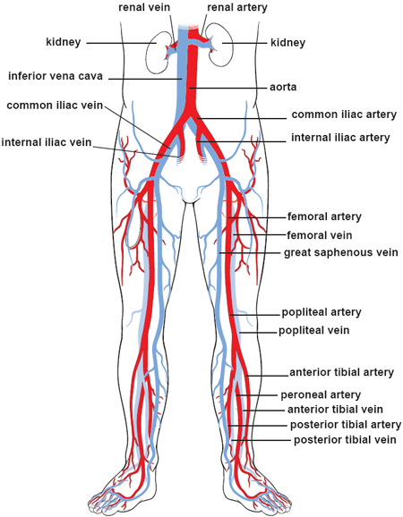
This full color stock medical exhibit illustrates the normal anatomy of the abdominal blood vessels.
Label the biliary passages and associated structures using the hints provided. In human anatomy, inferior epigastric artery refers to the artery that arises from the external iliac artery.it anastomoses with the superior epigastric artery.along its course, it is accompanied by a similarly named vein, the inferior epigastric vein.these epigastric vessels form the lateral border of hesselbach's triangle, which outlines the area through which direct inguinal hernias protrude. Aortas or aortae 4) is the main blood vessel in the abdominal cavity that transmits oxygenated blood from the thoracic cavity to the organs within the abdomen and to the lower limbs. Katy wallis at state college of florida Arteries (in red) are the blood vessels that deliver blood to the body. The venous drainage of the abdomen is carried out by the portal venous system and the systemic venous system. Doppler studies of the abdominal vessels demand an understanding of normal and abnormal blood flow patterns. Learn vocabulary, terms, and more with flashcards, games, and other study tools. Most of this blood is shunted into the aortic arch through the ductus arteriosus. The superior vena cava receives blood from all areas above the diaphragm (with the heart wall as the exception). The initial articles provided an overview of basic ultrasonography principles and a discussion about how to perform a systematic scan of the abdomen. Deep veins, located in the center of the leg near the leg bones, are enclosed by muscle. We undertook a review of the anatomical changes of choke vessels between the internal thoracic artery (ita) and deep inferior epigastric artery (diea), as highlighted by a case of aortoiliac occlusive disease (leriche's syndrome), and discuss the physiological concepts observed with regard to surgical delay procedures within the abdominal wall performed prior to abdominal cutaneous free.
Ventral view of heart with ventricle pulled anteriorly to expose part of sinus venosus. The videos are done by dr. Dogfish shark heart & associated branchial blood vessels. The identification of abdominal vessels using ultrasound is based on knowledge of their normal location, appearance and relationship to specific organs. Nerves and vessels branches derived from the descending aorta supply the posterior abdominal wall.
Illustrations Of The Blood Vessels from my.clevelandclinic.org Arteries (in red) are the blood vessels that deliver blood to the body. Read the other small animal abdominal ultrasonography articles. The aorta is the largest blood vessel in the body. Courses inferior through chest and enters abdomen through the diaphragm. Ventral view of heart with ventricle pulled anteriorly to expose part of sinus venosus. Nerves, blood vessels, and lymphatics are present throughout. It is an artery, meaning that it carries blood away from the heart. The rest of the series discusses ultrasound evaluation of specific abdominal organs/systems.
The abdominal aorta supplies blood to much of the abdominal cavity.
Abdominal wall anatomy that is clinically pertinent to the surgeon, focusing primarily on the structures of the anterior abdominal wall, will be reviewed. Ventral view of heart with ventricle pulled anteriorly to expose part of sinus venosus. Courses inferior through chest and enters abdomen through the diaphragm. Label the biliary passages and associated structures using the hints provided. The videos are done by dr. The abdominal aorta enters the abdomen through the diaphragm at the level of the twelfth thoracic vertebre and continues to just below the umbilical area, where it splits into the right and left common iliac arteries. Katy wallis at state college of florida The rest of the series discusses ultrasound evaluation of specific abdominal organs/systems. The venous drainage of the abdomen is carried out by the portal venous system and the systemic venous system. The abdominal aorta is clinically divided into 2 segments: This full color stock medical exhibit illustrates the normal anatomy of the abdominal blood vessels. Thoracic & abdominal blood vessels eeob 235: Label the intestinal structures using the hints provided.


0 Komentar